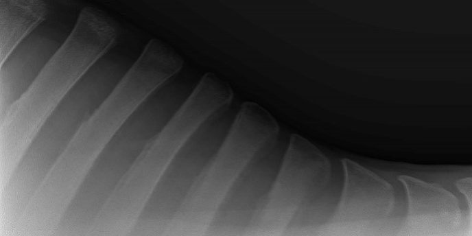
This is the name given not to “affectionate backs” but to over-riding or impinging of the dorsal spinous processes of the vertebrae commonly in thoracic (chest) or lumbar (lower back) region of the horse.
Often it is in the region of wither or saddle and can be associated with a poor saddle fit or trauma/damage often from as far back as when the horse was being broken in or weaning. Horses that rear up and over backwards and land on their withers is a common “accident” that can cause injury in this area. Sometimes we’ll never know the cause.
The damage or pressure can involve ligaments /fractures or callus formation leading to pinching or impinging of the space between bony finger like projections.
It is important to realise that radiographic changes can be apparent but the horse may be free from back pain which can be problematic at times such as pre purchase time when we have to assess risks.
Radiographic changes on their own or clinical signs on their own are not definitive of the condition.
Clinically the condition may be suspected with horses that are cold backed/have a poor action when saddled up/or a poor canter action.
On palpation they may show muscle spasm and dorsal back pain often localising into specific vertebrae.
Obviously, all bad signs for ridden exercise in any discipline!!!
Radiography is only possible laterally (from the side) due to the large mass of the back muscles and has limitations with only being able to visualise the upper half of vertebrae with this view. Ultrasound can be used to investigate ligament attachments/articulations.
Referral for ultrasound or bone scan are other options but a trial medication with a local anaesthetic and /or cortisone may be required to assess relevance also. Sometimes a big change on a radiograph causes little or no pain and a small change on a radiograph causes a lot of discomfort.
In summary- diagnosis and management of this condition is based on clinical signs + radiographic changes. Sometimes response to treatment helps reach a diagnosis as well as nerve blocking the area.
Treatment or management is based around cortisone injections (repeat injections often required)–possibly shockwave treatments- massage/hot packs- in extreme cases surgical resection may be tried. Recovery from surgery can be up to six months.
If you suspect a case of this then consult your favourite vet for a work up of clinical signs/x-rays /nerve blocks/cortisone injections if required.


