Dave Kruger, Vet Services Napier
“Boy” is an eight year old pony who was presented for a routine dental examination/float.
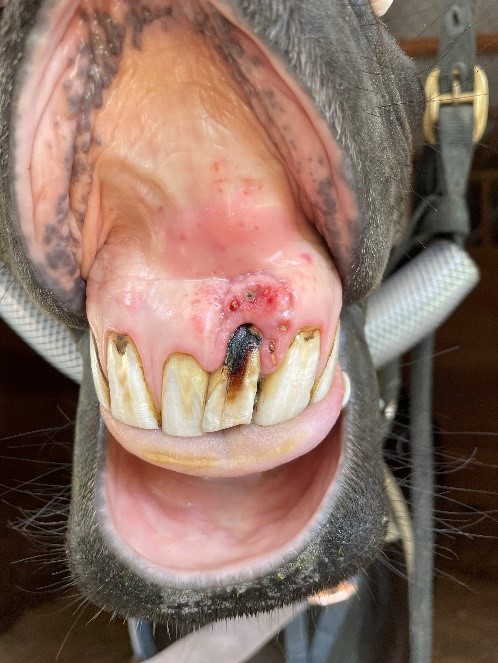
During the examination Dave Kruger found his central upper left incisor (tooth number 201 – modified Triadan system) was notably discoloured with some gingival (gum) loss at the tooth margin. A small draining tract was evident just above the tooth indicating a chronic tooth root abscess. The tooth was not loose on palpation.
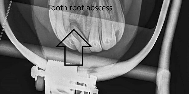
X-rays revealed significant shortening of the reserve crown of this tooth (compared to adjacent central upper right incisor) with an area of bone resorption at the root tip confirming a tooth root abscess. At this point the decision was made to extract the tooth.
After sedation a local infraorbital nerve block was performed. This involves injecting local anaesthetic into the infraorbital canal in order to block the upper arcade of teeth.
Regional local anaesthetic was also injected.
After incision of the edge of the gingiva (gum) around the tooth margin a periodontal elevator and incisor spreader was used to gently loosen the tooth within it’s socket. The aim with any tooth extraction is to very patiently loosen the tooth until the periodontal membrane (the structure which hold the tooth in place) is broken down and the tooth can be lifted out without applying any undue force.
Post operatively pain killers and antibiotics were prescribed for a week.
There is no practical way to close the gum after extraction in a horse and the wound was left to heal by “second intention” healing.
Because horses teeth erupt at a constant rate throughout their lives, it is vital that annual dental check are performed after any extraction. The opposing tooth has nothing to wear against and needs to be reduced to prevent step formation and potential restriction of the normal chewing/biting action.
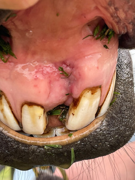
6 week post operative – healthy granulation tissue filing defect.


